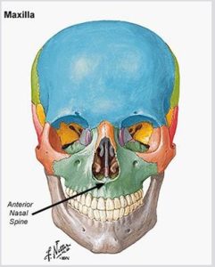
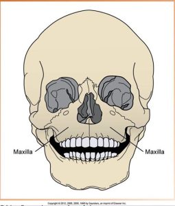
Paired bones of the maxilla
The upper jaw is composed of two paired bones, the maxillae. The paired maxillae meet at the midline of the face. The paired maxillae are often referred to as one single bone, the maxilla. All of the bones in the face articulate with the maxilla. The lower floor of the maxilla supports the upper teeth. The upper floor forms the floor of the orbit of the eye, the sides and floor of the nasal cavities, and the hard palate.
Bony Landmarks of the Maxilla
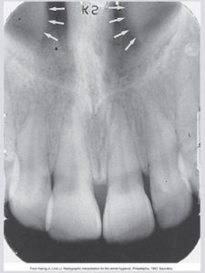
Incisive Foramen (Nasopalatine Foramen)
An opening or hole in bone that is located at the midline of the anterior portion of the hard palate directly posterior to the maxillary central incisors. The nasopalatine nerve exits the maxilla through the incisive foramen. It appears as a small ovoid or round radiolucent area located between the roots of the maxillary central incisors. The term foramen refers to a hole.
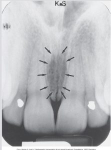
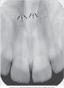
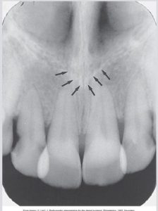
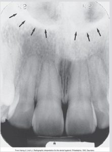
Superior Foramina of the Incisive Canal
Two tiny openings or holes in bone that are located on the floor of the nasal cavity. These join together to form the incisive canal. The superior foramina of the incisive canal appear as two small, round radiolucencies located superior to the apices of the maxillary central incisors.
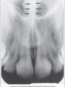
Median Palatal Suture
The immovable joint between the two palatine processes of the maxilla. A thin radiolucent line between the maxillary central incisors. The median palatine suture extends from the alveolar bone between the maxillary central incisors to the posterior hard palate.
Lateral fossa
The lateral fossa, also known as the canine fossa, is a smooth, depressed area of the maxilla located just inferior and medial to the infraorbital foramen between the canine and lateral incisors. The lateral fossa appears as a radiolucent area between the maxillary canine and lateral incisors. The appearance on the lateral fossa depends on the patient’s individual anatomy.
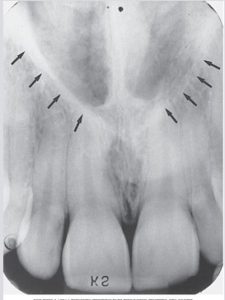
Nasal Cavity or Nasal Fossa
The nasal cavity, also known as the nasal fossa, is a pear-shaped compartment of bone located superior to the maxilla. The inferior portion is formed by the palatal processes of the maxilla and the horizontal portions of the palatine bones. The lateral walls of the nasal cavity are formed by the ethmoid bone and the maxillae. The nasal cavity appears as a large radiolucent area above the maxillary incisors. The nasal cavity is divided by the nasal septum.
Nasal Septum
A vertical bony wall or partition that divides the nasal cavity into the right and left nasal fossae. Formed by the vomer and a portion of the ethmoid bone and cartilage. The nasal septum appears as a vertical radiopaque partition that divides the nasal cavity.
Floor of the Nasal Cavity
A bony wall formed by the palatal processes of the maxilla and the horizontal portions of the palatine bones. The floor is composed of dense cortical bone and defines the inferior border of the nasal cavity. The floor of the nasal cavity appears as a dense radiopaque band of bone above the maxillary incisors.
Anterior Nasal Spine
A sharp projection of the maxilla located at the anterior and inferior portion of the nasal cavity. The anterior nasal spine appears as a V-shaped radiopaque area located at the intersection of the floor of the nasal cavity and the nasal septum.
Inferior Nasal Concha
Wafer-thin, curved plates of bone that extend from the lateral walls of the nasal cavity. Conchae means shell-shaped or scroll-shaped. The inferior nasal conchae appears as s diffuse radiopaque mass or projection within the nasal cavity.
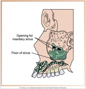
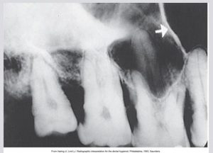
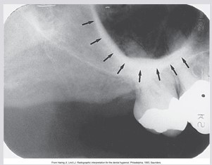
Maxillary Sinus
The maxillary sinuses are paired cavities or compartments of bone located within the maxilla, above the maxillary premolar and molar teeth. At birth, the maxillary sinus is the size of a small pea. The maxillary sinus appears as a radiolucent area located above the apices of the maxillary premolars and molars. The floor of the maxillary sinus is composed of dense cortical bone.
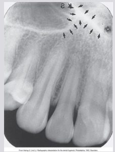
Septa within the Maxillary Sinus
Bony walls or partitions that appear to divide the maxillary sinus into compartments. Septa is plural for septum. The septa appears as radiopaque lines within the maxillary sinus. Presence and number vary depending on the anatomy of the individual.
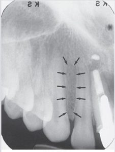
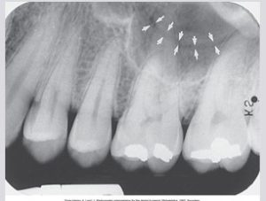
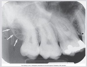
Nutrient Canals within the Maxillary Sinus
Tiny, tubelike passageways through bone that contain blood vessels and nerves, supplying the maxillary teeth and interdental areas. The nutrient canals appear as a narrow radiolucent band bounded by two thin radiopaque lines. The radiopaque lines represent the cortical bone that makes up the walls of the canal.
Inverted Y
The intersection of the maxillary sinus and the nasal cavity. The inverted Y appears as a radiopaque upside-down Y formed by the intersection of the lateral wall of the nasal fossa and the anterior border of the maxillary sinus. Located above the maxillary canine, both the lateral wall of the nasal cavity and the anterior border of the maxillary sinus are composed of dense cortical bone.
Maxillary Tuberosity
A rounded prominence of bone that extends posterior to the third molar region. Blood vessels and nerves enter the maxilla in this region and supply the posterior teeth. The maxillary tuberosity appears as a radiopaque bulge distal to the third molar region.
Hamulus
A small hooklike projection of bone extending from the medial pterygoid plate of the sphenoid bone. This is also known as the hamular process. The hamulus is located posterior to the maxillary tuberosity region. The hamulus appears as a hooklike radiopaque projection posterior to the maxillary tuberosity area. The radiographic appearance of the hamulus varies in length, shape, and density.
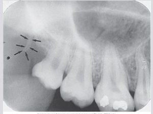
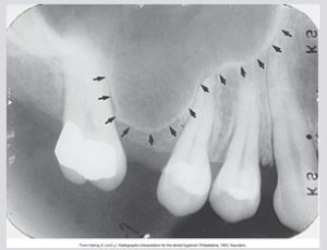
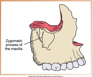
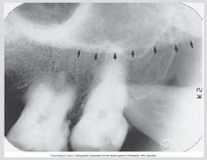
Zygomatic Process of the Maxilla
A bony projection of the maxilla that articulates with the zygoma or malar (cheek) bone. The zygomatic process of the maxilla appears as a J-shaped or U-shaped radiopacity superior to the maxillary first molar region. The zygomatic process of the maxilla is composed of dense cortical bone.
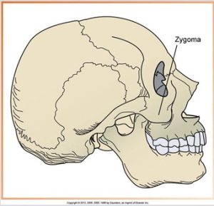
Zygoma
The zygoma, also known as the cheekbone, malar bone, or zygomatic bone, articulates with the zygomatic process of the maxilla. The zygoma is composed of dense cortical bone, and appears as a diffuse radiopaque band extending posteriorly from the zygomatic process of the maxilla.