25.2
Diagnostic Panoramic Image
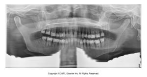
Diagnostic panoramic images are the results when the equipment preparation, patient preparation, and patient positioning are completed correctly. They should ideally be free from all errors and must demonstrate accurate anatomic features and proper exposure, resulting in correct density and contrast.
Below is an image is a panoramic dental x-ray showing the upper and lower jaws, teeth, and sinuses.
Anatomic Features
Panoramic images must allow for the visualization of the maxillofacial anatomic features that it represents. Each panoramic image should be assessed to determine if the dentition and bones of the maxillofacial region are representative.
Density and Contrast
Diagnostic panoramic images result from adequate exposure and exhibit proper density and contrast. The smaller patients require less exposure. With an ideal panoramic image, the density (overall darkness) is not excessive. Proper contrast on a panoramic image is critical and should allow for identification of the junction between enamel and dentin in the molar region.
Below are images of panoramic dental x-rays with exposures showing detailed structures of adult teeth, jawbones, and sinus areas.
Patient Preparation Errors
Patient preparation errors include ghost images and lead apron artifacts.
Ghost Images
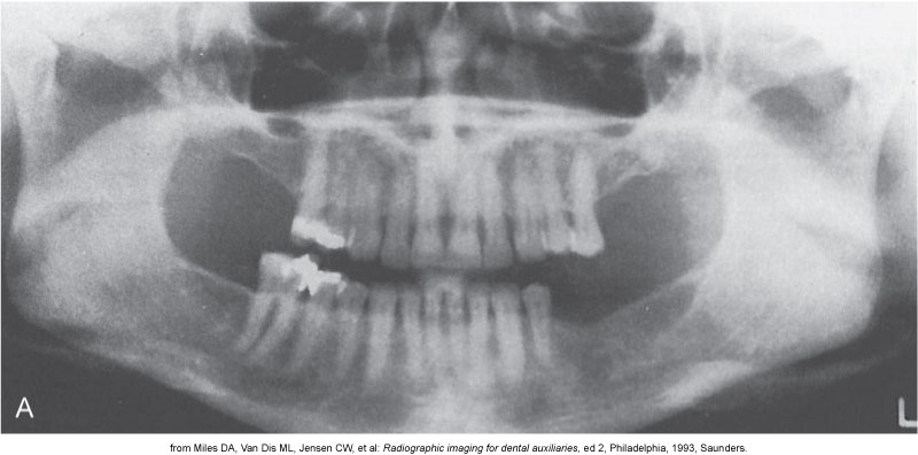
Ghost images are when an imaging artifact is seen on a panoramic image that is produced when a metallic or dense object is penetrated twice by the x-ray beam.
It is found on the opposite side of the receptor, and it appears indistinct, larger, and higher than its actual counterpart.
Problem: If all metallic or radiodense objects are not removed before exposure, a ghost image results that obscures diagnostic information.
Solution: The dental radiographer must instruct the patient to remove all radiodense objects in the head and neck region before positioning the patient.
See an example of a ghost image below.
Lead Apron Artifacts
Problem: A radiopaque cone-shaped artifact that obscures diagnostic information results if the lead apron is incorrectly placed, or if a lead apron with a thyroid collar is used.
Solution: The dental radiographer must always use a lead apron without a thyroid collar when exposing a panoramic projection.
See an example of a lead apron artifact image below.
Patient Positioning Errors
The different patient positioning errors include:
- Positioning of the lips and tongue
- Chin tipped up
- Chin tipped down
- Teeth anterior to the focal trough
- Teeth posterior to the focal trough
- Head turned
- Slumped posture
Go through the slides below to see examples, problems, and solutions for each of these errors.
Advantages & Disadvantages of Panoramic Radiography
| Advantages: | Disadvantages: |
|
|
Media Attributions
- Iannuci: Dental Radiography, 6th Edition, Chapter 25, CC BY-NC-ND
