2.3
Production of Dental X-Rays
- Electricity from the wall outlet supplies the power to generate x-rays.
- The current is directed to the filament circuit and step-down transformer in the tubehead.
- The filament circuit uses 3 to 5 volts to heat the tungsten filament in the cathode.
- Thermionic emission occurs, and the release of electrons. The electrons stay in an electron cloud until the high-voltage circuit is activated.
- When the exposure button is pushed, the high-voltage circuit is activated.
- The molybdenum cup in the cathode directs electrons to the tungsten target and into the anode.
- When electrons strike the tungsten target, less than 1% of the energy is converted to
x-rays, and the remaining 99% is lost as heat. - A small number of x-rays are able to exit the x-ray tube through the unleaded glass window portion of the tube.
- X-rays travel through the unleaded glass window, the tubehead seal, and aluminum disks.
- The size of the x-ray beam is restricted by the lead collimator.
- The x-ray beam exits the tubehead at the opening of the PID.
Types of X-Rays Produced
X-rays differ in energy and wavelength. Two mechanisms produced are general (braking) radiation and characteristic radiation.
General Radiation (AKA: Braking radiation (bremsstrahlung)
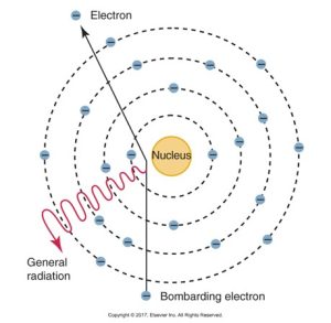
Many electrons that interact with the tungsten atoms undergo not one but many interactions within the target. Most x-rays (about 70%) are produced in this manner and are reduced when an electron hits the nucleus of a tungsten atom or passes very close to the nucleus of a tungsten atom.
General radiation occurs when an electron that passes close to the nucleus of a tungsten atom is slowed down; an x-ray photon of lower energy results.
The diagram below shows the interaction between a bombarding electron and an atom, resulting in the release of general radiation, depicted as waves emanating from the point of collision.
Characteristic Radiation
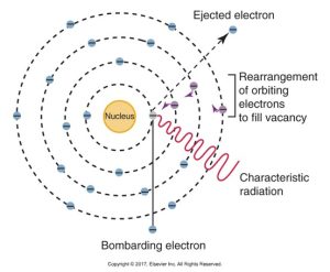
Characteristic radiation is produced when a high-speed electron dislodges an inner-shell electron from a tungsten atom and causes ionization of that atom. The remaining orbiting electrons are rearranged to fill the vacancy. This occurs only at 70 kVp and above.
Characteristic radiation is produced when an electron dislodges an inner-shell electron from the tungsten atom, resulting in the rearrangement of remaining orbiting electrons.
The diagram below illustrates characteristic radiation in an atom, showing a bombarding electron causing the ejection of an orbital electron and subsequent rearrangement of electrons to fill the vacancy, resulting in the emission of characteristic radiation.
Definitions of X-Radiation
Interactions of X-Radiation
When X-rays pass through matter, several outcomes are possible:
- There can be no interaction, with X-rays traveling through without any change.
- The absorption of energy and the photoelectric effect may occur; this is when an X-ray photon is fully absorbed, resulting in the ejection of an electron from an inner shell.
- Compton scatter can happen, where an X-ray photon collides with an outer shell electron, scattering the photon and ejecting the electron.
- Coherent scatter takes place when an X-ray photon causes a temporary excitation in an atom, leading to the photon being scattered without a loss of energy.
There are three types of radiation interactions with the patient that may occur:
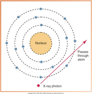
No Interaction
No interaction can happen when the x-ray photon passes through the atom unchanged and leaves the atom unchanged. These photons are responsible for producing densities on film and make dental radiography possible.
The diagram below illustrates an x-ray photon passing through an atom without interaction, showing the photon’s trajectory as it moves straight past the atomic nucleus and electron shells.
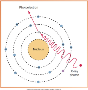
Absorption of Energy and Photoelectric Effect
The x-ray photon is completely absorbed within matter or the tissues of the patient.
Absorption is the total transfer of energy from photons to the atoms of matter, and the Photoelectric Effect is when an x-ray photon collides with a tightly bound, inner-shell electron and gives up all its energy to eject the electron from its orbit. The ejected photoelectron is absorbed by other atoms.
When an x-ray photon collides with an inner-shell electron, a photoelectric effect occurs: The photon is absorbed and ceases to exist, and a photoelectron with a negative charge is produced.
The diagram below depicts the photoelectric effect, where an x-ray photon collides with an atom, resulting in the ejection of a photoelectron from the electron shell.
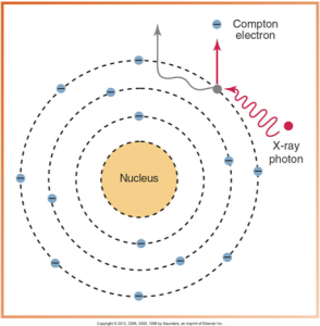
Compton Scatter
The x-ray photon is deflected from its path during its passage through matter. In Compton scatter, ionization takes place and an x-ray photon collides with a loosely bound, outer-shell electron and gives up part of its energy to eject the electron from its orbit. The x-ray photon loses energy and continues in a different direction at a lower energy level. The ejected electron is termed a Compton electron.
Compton or Recoil Electron
When an x-ray photon collides with an outer-shell electron and ejects the electron from its orbit, Compton scatter results: The photon is scattered in a different direction at a lower energy, and the ejected electron is referred to as a Compton, or recoil, electron.
The diagram below illustrates the compton effect, with an x-ray photon colliding with an outer electron and causing the electron’s ejection as a compton electron while the photon changes direction.
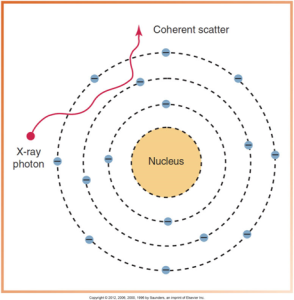
Coherent Scatter
Coherent scatter is an x-ray photon that has its path altered by matter. A low-energy x-ray photon interacts with an outer-shell electron, and no change in the atom occurs. An x-ray photon of scatter radiation is produced.
Coherent Effect
The coherent effect is when an x-ray photon is scattered, and no loss of energy occurs; the scatter is termed coherent.
The diagram below illustrates a coherent scatter, depicting an x-ray photon that changes direction without a change in energy after interacting with an atom’s electron cloud.
Media Attributions
- Iannucci & Howerton: Dental Radiography Principles and Techniques, 6th Edition, Chapter 2, CC BY-NC-ND
