9.1 DNA Structure and Replication
Discovery of DNA
Determining that DNA is the genetic material on an organism was an important milestone in biology. It took many scientists undertaking creative experiments over several decades to show with certainty that DNA is the molecule that determines the traits of organisms.
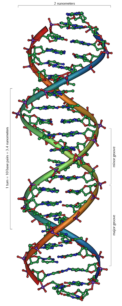
One of the most significant scientific achievements of the 20th century was the discovery of the DNA double helix, a twisted ladder-like structure. In the 1950s, James Watson and Francis Crick, with crucial contributions from Rosalind Franklin and Maurice Wilkins, unveiled the double helix model of DNA (see Figure 9.1.1).
Figure 9.1.1 Description
3D molecular model of the DNA double helix, showing its twisted ladder-like structure. The image highlights the major and minor grooves, with a full turn of the helix measuring 3.4 nanometers and 10 base pairs per turn. The diameter of the helix is labelled as 2 nanometers.
Building Blocks of DNA
The building blocks of deoxyribonucleic acid (DNA) are nucleotides, which are made up of three parts: a deoxyribose (5-carbon sugar), a phosphate group, and a nitrogenous base (Figure 9.1.2).
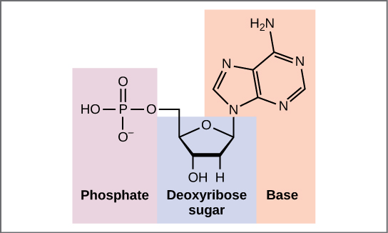
Figure 9.1.2 Description
Diagram of a nucleotide showing its three main components: a phosphate group, a ribose sugar, and a nitrogenous base. The phosphate group is highlighted in purple, the ribose sugar in blue, and the nitrogenous base in orange.
There are four types of nitrogenous bases in DNA. Adenine (A) and guanine (G) are double-ringed purines, and cytosine (C) and thymine (T) are smaller, single-ringed pyrimidines. The nucleotide is named according to the nitrogenous base it contains.
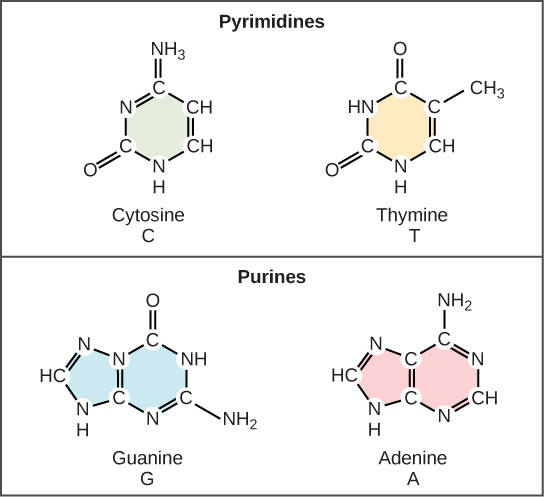
Figure 9.1.3 Description
Diagram showing the chemical structures of the four nitrogenous bases in DNA, categorized into two groups: pyrimidines (cytosine and thymine) and purines (guanine and adenine). Pyrimidines have a single-ring structure, while purines have a double-ring structure. Each base is labelled with its name, abbreviation, and coloured background.
Structure of DNA
The phosphate group of one nucleotide bonds covalently with the sugar molecule of the next nucleotide, and so on, forming a long polymer of nucleotide monomers. The sugar-phosphate groups line up in a “backbone” for every single strand of DNA, and the nitrogenous bases stick out from this backbone. The carbon atoms of the five-carbon sugar are numbered clockwise from the oxygen as 1′, 2′, 3′, 4′, and 5′ (1′ is read as “one prime”). The phosphate group is attached to the 5′ carbon of one nucleotide and the 3′ carbon of the next nucleotide. In its natural state, each DNA molecule is actually composed of two single strands held together along their length with hydrogen bonds between the bases.
DNA comprises two strands twisted around each other to form a helix, called a double helix. Base pairing takes place between a purine and pyrimidine: namely, A pairs with T, and G pairs with C. In other words, adenine and thymine are complementary base pairs, and cytosine and guanine are complementary base pairs. The two strands are anti-parallel in nature; that is, one strand will have the 3′ carbon of the sugar in the “upward” position, whereas the other strand will have the 5′ carbon in the upward position. The diameter of the DNA double helix is uniform throughout because a purine (two rings) always pairs with a pyrimidine (one ring), and their combined lengths are always equal. (Figure 9.1.3).

Figure 9.1.4 Description
Illustration of DNA structure. Panel (a) shows a DNA double helix with base pairs colored by type: adenine (red), thymine (yellow), guanine (blue), and cytosine (green), along with the sugar-phosphate backbone. Panel (b) shows a zoomed-in view of DNA base pairing: adenine pairs with thymine via two hydrogen bonds, and guanine pairs with cytosine via three hydrogen bonds. Sugar-phosphate backbones are shown on both sides of the base pairs with directionality labelled 5′ to 3′.
Exercises 9.1.1
Text Description
1. Two-ring bases always bind to each other (True/False?)
2. In DNA, each nucleotide contains a sugar. (True/False?)
3. Complementary Base Pairing- drag and drop to complete the complementary strand
Drop Zones:
- A
- C
- G
- T
- C
- A
Possible Answers:
- A
- T
- C
- G
4. A single strand of DNA is a polymer of ____ joined ____ between the ____ of one and the ____ of the next to form a “backbone” from which the ____ bases stick out. In its natural state, DNA has ____ wound around each other in a ____. The bases on each strand are bonded to each other with ____ bonds. Only specific bases bond with each other; ____ bonds with ____, and ____ bonds with ____.
Answers
- False
- True
- A: T
C: G
G: C
T: A
C: G
A: T - A single strand of DNA is a polymer of nucleic acids joined covalently between the phosphate group of one and the deoxyribose sugar of the next to form a “backbone” from which the nitrogenous bases stick out. In its natural state, DNA has two strands wound around each other in a double helix. The bases on each strand are bonded to each other with hydrogen bonds. Only specific bases bond with each other; adenine bonds with thymine, and cytosine bonds with guanine.
DNA Replication
DNA replication is required for the growth or replication of an organism. You started as one single cell and are now made up of approximately 37 trillion cells! Each and every one of these cells contains the exact same copy of DNA, which is only possible because of DNA replication.
DNA replication is the process by which DNA is copied. It occurs during the synthesis (S) phase of the eukaryotic cell cycle. DNA must be copied so that each new daughter cell will have a complete set of chromosomes after cell division occurs.

Figure 9.1.5 Description
Diagram of DNA replication showing the unwinding of the double helix into two single strands. Each strand serves as a template for synthesizing a new complementary strand. Arrows indicate the direction of replication, and base pairing is shown between the template and new strands.
Knowledge of DNA’s structure helped scientists understand how DNA is copied. Recall that adenine nucleotides pair with thymine nucleotides and cytosine with guanine. This means that the two strands are complementary to each other. For example, a strand of DNA with a nucleotide sequence of AGTCATGA will have a complementary strand with the sequence TCAGTACT.
DNA replication is referred to as semi-conservative. This means that when a strand of DNA is replicated, each of the two original strands acts as a template for a new complementary strand. When the replication process is complete, there are two identical sets of DNA, each containing one of the original DNA strands and one newly synthesized strand.
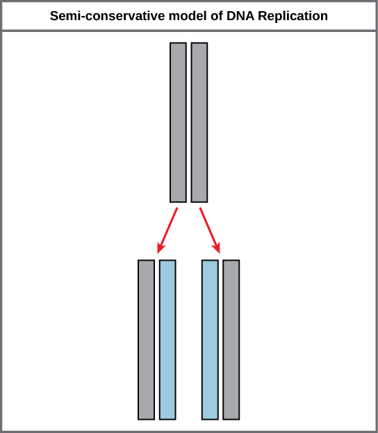
Figure 9.1.6 Description
Diagram illustrating the semi-conservative model of DNA replication. The original DNA molecule, shown as two gray strands, separates, and each strand serves as a template for a new strand. The resulting DNA molecules each contain one original (gray) strand and one newly synthesized (blue) strand.
DNA replication is a complicated process with a specific sequence of events and enzymes that facilitate each step. DNA polymerases are key enzymes that form covalent bonds between nucleotides to build a new DNA strand. Replication starts at specific sites called origins of replication, where the parental DNA strands start to unwind in both directions, forming replication bubbles. Once the nitrogenous bases from the inside of the parental DNA molecule are exposed, the creation of a new, complementary strand can begin. As each nucleotide pairs with its complementary base on the template strand, DNA polymerase creates a covalent bond to attach to the growing daughter strand. Eventually, all replication bubbles merge, resulting in two complete double-stranded daughter DNA molecules.
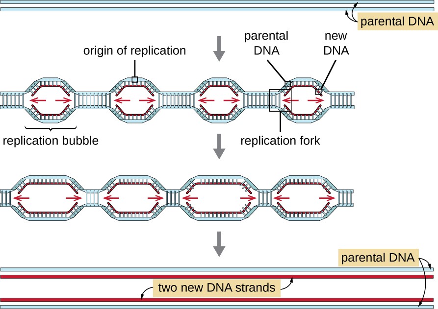
Figure 9.1.7 Description
A diagram showing two strands of parental DNA. Then, an arrow shows multiple replication bubbles with an origin of replication in each. Arrows point to the left and right from each origin of replication. New strands of DNA are shown to be formed. One of the bubbles has the left half of the bubble in a box labelled replication fork. The next image shows the replication bubbles getting longer. The final image shows two new DNA strands, each with one old strand and one new strand.
“5.3 DNA” from Human Biology by Christine Miller is licensed under a Creative Commons Attribution-NonCommercial 4.0 International License, except where otherwise noted.
“9.1 The Structure of DNA” from Biology and the Citizen by Colleen Jones is licensed under a Creative Commons Attribution 4.0 International License, except where otherwise noted.
“5.4 DNA Replication” from Human Biology by Christine Miller is licensed under a Creative Commons Attribution-NonCommercial 4.0 International License, except where otherwise noted.
“9.2 DNA Replication” from Biology and the Citizen by Colleen Jones is licensed under a Creative Commons Attribution 4.0 International License, except where otherwise noted.
