7.4 Meiosis
Sexual Reproduction
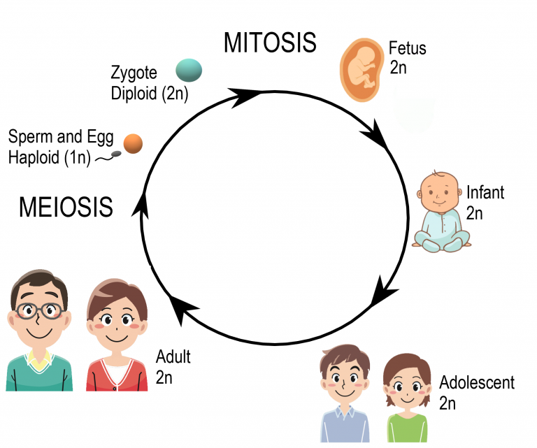
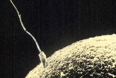
Reproduction is the process by which organisms give rise to offspring. It is one of the defining characteristics of living things. Like many other organisms, human beings reproduce sexually. Sexual reproduction involves two parents. As you can see from Figure 7.4.1, in sexual reproduction, parents produce reproductive (sex) cells — called gametes — that unite to form offspring. Gametes are haploid (or n) cells. This means they contain one copy of each chromosome in the nucleus. Gametes are produced by a type of cell division called meiosis, which is described in detail below. The process in which two gametes unite is called fertilization. The fertilized cell that results is referred to as a zygote. A zygote is a diploid (or 2n) cell which contains two copies of each chromosome. Thus, it has twice the number of chromosomes as a gamete.
Meiosis
The process that produces haploid gametes is called meiosis. Meiosis is a type of cell division in which the number of chromosomes is reduced by half. During meiosis, homologous chromosomes (paired chromosomes — one from each parent) separate in the first division (meiosis I), so the resulting cells only have one chromosome from each pair. These cells then go through a second division (meiosis II), where the sister chromatids of each chromosome are then separated, ensuring that each gamete ends up with a single copy of each chromosome.
This type of cell division starts with one diploid cell and finishes with four genetically different haploid cells. Meiosis is only used to produce gametes for sexual reproduction.
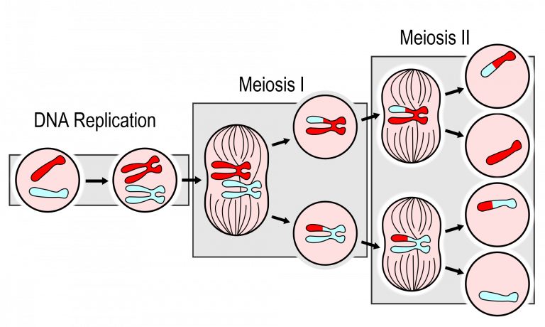
As you can see in the meiosis diagram, the two cell divisions are called meiosis I and meiosis II. Meiosis I begins after DNA replicates during interphase. Meiosis II follows meiosis I without DNA replicating again. Both meiosis I and meiosis II occur in four phases: prophase, metaphase, anaphase, and telophase. You may recognize these four phases from mitosis, the division of the nucleus that takes place during routine cell division of eukaryotic cells.
Meiosis I- Increasing Genetic Variation
The phases of Meiosis I are:
- Prophase I: The nuclear envelope begins to break down, and the chromosomes condense. Centrosomes start moving to opposite cell poles, and spindle fibers form. Importantly, homologous chromosomes pair up, which is unique to prophase I. In the prophase of mitosis and meiosis II, homologous chromosomes do not form pairs in this way. During prophase I, crossing-over occurs. The significance of crossing over is discussed below.
- Metaphase I: Spindle fibres attach to the paired homologous chromosomes. The paired chromosomes line up along the cell’s equator, randomly aligning in a process called independent assortment (discussed below). This occurs only in metaphase I. In the metaphase of mitosis and meiosis II, sister chromatids line up along the cell’s equator.
- Anaphase I: Spindle fibres shorten, and the chromosomes of each homologous pair start to separate from each other. One chromosome of each pair moves toward one pole of the cell, and the other chromosome moves toward the opposite pole.
- Telophase I and Cytokinesis: The spindle breaks down, and new nuclear membranes form. The cytoplasm of the cell divides, and two haploid daughter cells result. The daughter cells each have a random assortment of chromosomes, with one from each homologous pair. Both daughter cells go on to meiosis II.
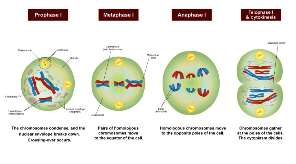
Figure 7.4.3 Meiosis I is critical in creating genetic diversity in resulting gametes. Crossing over in Prophase I and independent alignment in Metaphase I ensure that each resulting gamete is unique. Image by Ali Zifan, CC BY-SA 4.0
Figure 7.4.3 Description
Diagram showing the stages of Meiosis I: Prophase I, Metaphase I, Anaphase I, and Telophase I with cytokinesis. In Prophase I, chromosomes condense, the nuclear envelope breaks down, and crossing-over occurs. In Metaphase I, homologous chromosomes align at the equator. In Anaphase I, homologous chromosomes move to opposite poles. In Telophase I and cytokinesis, chromosomes gather at the poles, and the cytoplasm divides, forming two cells.
Meiosis II- Halving the DNA
The phases of Meiosis II are:
- Prophase II: The nuclear envelope breaks down, and the spindle forms in each haploid daughter cell from meiosis I. The centrosomes also start to separate.
- Metaphase II: Spindle fibres line up the sister chromatids of each chromosome along the cell’s equator.
- Anaphase II: Sister chromatids separate and move to opposite poles.
- Telophase II and Cytokinesis: The spindle breaks down, forming new nuclear membranes. The cytoplasm of each cell divides, and four haploid cells result. Each cell has a unique combination of chromosomes.
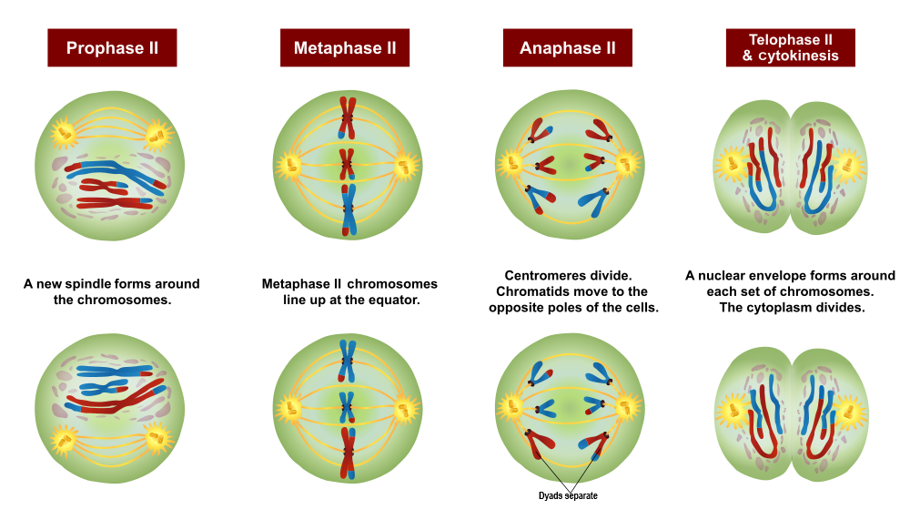
Figure 7.4.4 Description
Diagram showing the stages of Meiosis II: Prophase II, Metaphase II, Anaphase II, and Telophase II with cytokinesis. In Prophase II, a new spindle forms around the chromosomes. In Metaphase II, chromosomes align at the cell equator. In Anaphase II, centromeres divide, and chromatids move to opposite poles. In Telophase II and cytokinesis, nuclear envelopes form around each set of chromosomes, and the cytoplasm divides, resulting in four genetically unique haploid cells.
Comparing Mitosis and Meiosis
Mitosis and meiosis are both processes of nuclear division in eukaryotic cells. They have many similarities and distinct differences that lead to very different outcomes.
Mitosis involves a single division, resulting in two new cells. These cells are genetically identical to the original and have the same number of chromosome sets. For humans, mitosis starts with one diploid cell, resulting in two diploid cells. This type of cell division is used for growth and replacing dead or damaged cells. It can also be used for asexual reproduction in some organisms.
Meiosis, on the other hand, involves two divisions, resulting in four cells. These cells are genetically diverse and contain half the number of chromosomes. In humans, meiosis starts with one diploid cell and results in four haploid cells. This type of cell division is used to produce gametes for sexual reproduction.
The differences in outcomes between mitosis and meiosis are primarily a result of the behaviour of chromosomes in meiosis I. In mitosis, chromosomes line up along the metaphase plate individually (or “single file”). This configuration results in each chromosome getting split, with one sister chromatid going to either pole. In contrast, in meiosis I, homologous chromosome pairs bind together, crossover, and line up along the metaphase plate in tetrads (or “double file”). This configuration results in the tetrad splitting with one entire chromosome from each pair going to either pole (sister chromatids remain intact).
Meiosis II is more similar to mitosis. Chromosomes line up along the metaphase plate individually (or “single file”), dividing sister chromatids.
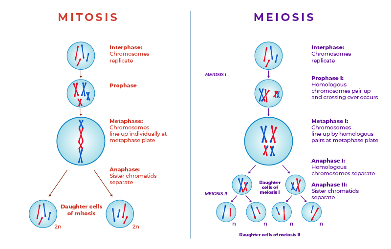
Figure 7.4.5 Description
Side-by-side comparison of Mitosis and Meiosis. On the left, the Mitosis process is shown: chromosomes replicate during interphase, align individually at the metaphase plate, and sister chromatids separate in anaphase, resulting in two diploid (2n) daughter cells. On the right, Meiosis is shown: chromosomes replicate during interphase, homologous chromosomes pair and undergo crossing over during Prophase I, align as pairs in Metaphase I, separate in Anaphase I, and then sister chromatids separate in Anaphase II. Meiosis results in four haploid (n) daughter cells, each genetically unique.
Sexual Reproduction and Genetic Variation
“It takes two to tango” might be a euphemism for sexual reproduction. Requiring two individuals to produce offspring, however, is also the main drawback of this way of reproducing because it requires extra steps — and often a certain amount of luck — to produce with a partner successfully. On the other hand, sexual reproduction greatly increases the potential for genetic variation in offspring, which increases the likelihood that the resulting offspring will have genetic advantages. In fact, each offspring produced is almost guaranteed to be genetically unique, differing from both parents and from any other offspring.
Sexual reproduction increases genetic variation in a number of ways:
Crossing Over
When homologous chromosomes pair up during meiosis I, crossing over can occur. Crossing over is the exchange of genetic material between non-sister chromatids of homologous chromosomes. It results in new combinations of genes on each chromosome.
Once crossing over has occurred, we can no longer call them sister chromatids since they are no longer identical; we term them dyads. In addition, once crossing over has occurred, the pair of homologous chromosomes can be referred to as tetrads.
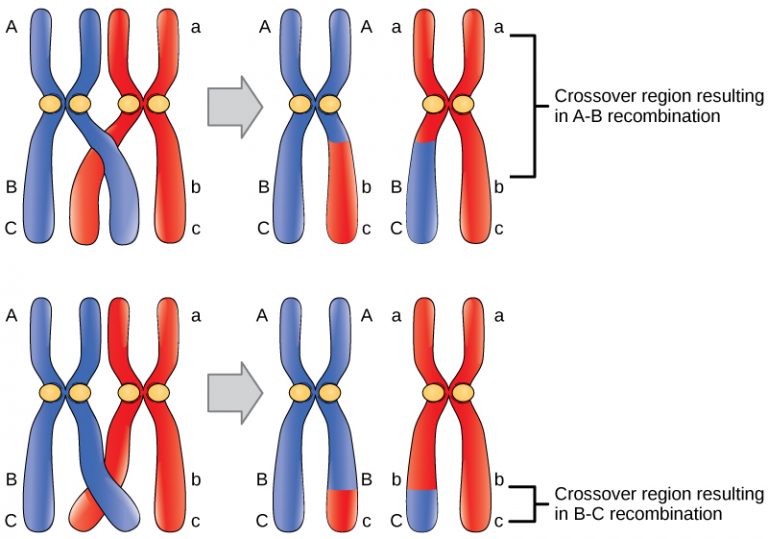
Figure 7.4.5 Description
Diagram showing crossing over between homologous chromosomes. The top panel shows A-B recombination, where a crossover occurs between regions labelled A and B on homologous chromosomes, resulting in an exchange of genetic material. The bottom panel shows B-C recombination, where the crossover occurs between regions B and C. Each results in chromosomes with mixed genetic segments, illustrating how recombination increases genetic diversity. Labelled regions A, B, C and their lowercase counterparts mark chromosome segments from different parents.
Independent Assortment
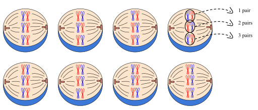
During metaphase I of meiosis, the orientation of each homologous pair of chromosomes is random. This means that either the maternal (red) or paternal (blue) chromosome can be on the left or right side. This is called independent assortment. It results in gametes that have unique combinations of chromosomes. For humans, independent assortment results in over 8 million different combinations of chromosomes!
For example, in Figure 7.4.7, there are 8 possible ways the chromosome pairs can align. In the first possibility, all 3 blue chromosomes are on the same side which would result in gametes with only red or only blue chromosomes. In the second possibility, the pairs are oriented differently, producing gametes with a mix of red and blue chromosomes. These different arrangements will result in gametes with different combinations of chromosomes.
Random Fertilization
In sexual reproduction, two gametes unite to produce an offspring. However, which two of the millions of potential gametes will combine is a matter of chance. Given that a human egg cell, with approximately 8 million possible genetic combinations, is randomly fertilized by a human sperm cell which also has about 8 million possibilities, a single man and woman can produce zygotes with 64 trillion combinations of chromosomes!
Errors in Meiosis
Mistakes may occur during meiosis that results in nondisjunction. This is the failure of replicated chromosomes to separate properly during meiosis. Some of the resulting gametes will be missing all or part of a chromosome, while others will have an extra copy of all or part of the chromosome. If such gametes are fertilized and form zygotes, they usually do not survive. If they do survive, the individuals are likely to have serious genetic disorders.
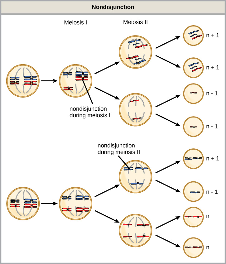
Figure 7.4.8 Description
Diagram illustrating nondisjunction during meiosis. In the top half, nondisjunction occurs during Meiosis I, where homologous chromosomes fail to separate, resulting in two gametes with an extra chromosome (n+1) and two with one missing chromosome (n–1). In the bottom half, nondisjunction occurs during Meiosis II, where sister chromatids fail to separate in one of the cells, producing one gamete with an extra chromosome (n+1), one with a missing chromosome (n–1), and two normal gametes (n).
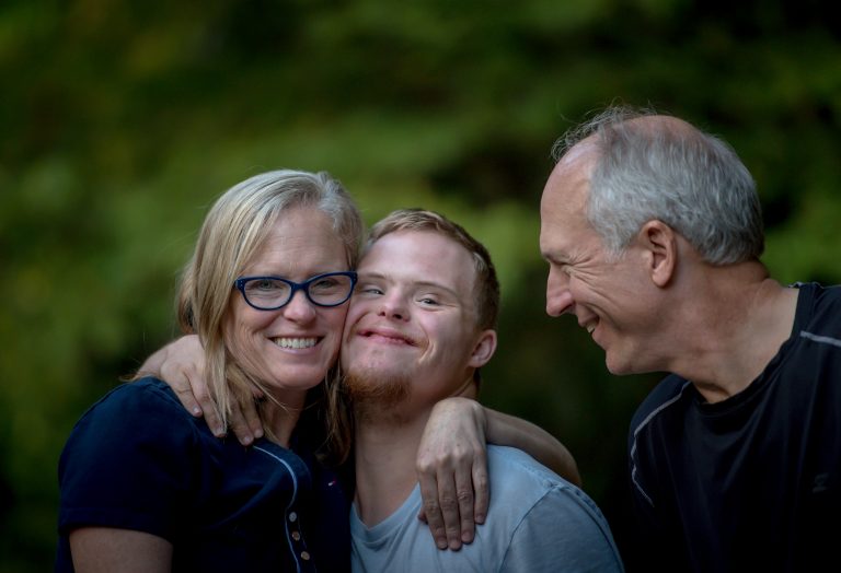
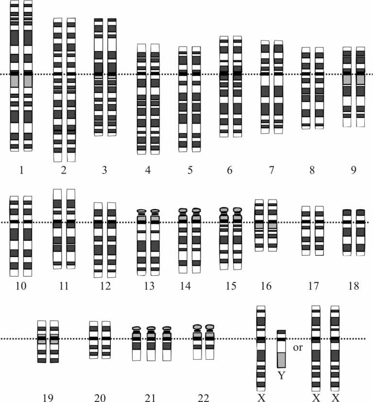
Down syndrome is a genetic disorder where an individual has an extra copy of chromosome 21. People with Down syndrome often have distinct facial features and developmental delays and may have other health issues.
Exercise 7.4.1
Text Description
In ____ crossing over occurs between homologous pairs of chromosomes.
In ____ independent alignment increases the genetic diversity that will occur once Meiosis is complete.
In ____ of mitosis, sister chromatids separate. In ____ of Meiosis, dyads separate. In ____ of Meiosis, tetrads separate.
Possible Answers:
- Anaphase I
- Prophase I
- Metaphase I
- Anaphase II
- Telophase I
- Anaphase
- Telophase II
Possible Drop Zones:
- Prophase 1
- Metaphase 1
- Anaphase 1
- Telophase 1
- Prophase 2
- Metaphase 2
- Anaphase 2
- Telophase 2
Possible Images:
- Image Illustrating Cells in Prophase 1
- Image Illustrating Cells in Metaphase 1
- Image Illustrating Cells in Anaphase 1
- Image Illustrating Cells in Telophase 1
- Image Illustrating Cells in Prophase 2
- Image Illustrating Cells in Metaphase 2
- Image Illustrating Cells in Anaphase 2
- Image Illustrating Cells in Telophase 2
5. How many daughter cells does meiosis produce?
- Four Haploids
- Two Haploids
- Two Diploids
- Four Diploids
6. At which stage of meiosis are sister chromatids separated from each other?
- Prophase I
- Anaphase II
- Prophase II
- Anaphase I
7. What is the part of meiosis that is similar to mitosis?
- Meiosis II
- Anaphase I
- Meiosis I
- Interkinesis
8. If a muscle cell of a typical organism has 32 chromosomes, how many chromosomes will be in a gamete of that same organism?
- 16
- 64
- 32
- 8
9. Drag the words into the correct boxes
Random alignment leads to new combinations of traits. The chromosomes that were originally inherited by the gamete-producing individual came equally from the ____ and the ____. In ____, the duplicated copies of these maternal and paternal homologous chromosomes line up across the ____ of the cell to form a tetrad. The ____ of each tetrad is ____. There is an ____ chance that the maternally derived chromosomes will be facing either pole. The same is true of the paternally derived chromosomes. The alignment should occur ____ in almost every meiosis. As the homologous chromosomes are pulled apart in ____, any combination of maternal and paternal chromosomes will move toward each pole. The gametes formed from these two groups of chromosomes will have a mixture of traits from the individual’s parents. Each gamete is ____.
Possible Answers:
- differently
- anaphase I
- egg
- sperm
- center
- orientation
- unique
- metaphase I
- random
- equal
10. Drag the words into the correct boxes to describe the ways meiosis II is similar to and different from mitosis of a diploid cell.
The two divisions are similar in that the chromosomes line up along the metaphase plate ____, meaning unpaired with other chromosomes (as in meiosis I). In addition, each chromosome consists of ____ sister ____ that will be pulled apart. The two divisions are different because in meiosis II there are ____ the number of chromosomes that are present in a diploid cell of the same species undergoing mitosis. This is because ____ reduced the number of chromosomes to a haploid state.
Possible Answers:
- half
- individually
- meiosis I
- chromatids
- two
3. In Prophase I crossing over occurs between homologous pairs of chromosomes.
In Metaphase I independent alignment increases the genetic diversity that will occur once Meiosis is complete.
In Anaphase of mitosis, sister chromatids separate. In Anaphase II of Meiosis, dyads separate. In Anaphase I of Meiosis, tetrads separate.
At the end of Telophase I there are two cells, and at the end of Telophase II there are four unique haploid cells.
6. Anaphase II
7. Meiosis II
8. Meiosis II
9. Random alignment leads to new combinations of traits. The chromosomes that were originally inherited by the gamete-producing individual came equally from the egg and the sperm. In metaphase I, the duplicated copies of these maternal and paternal homologous chromosomes line up across the center of the cell to form a tetrad. The orientation of each tetrad is random. There is an equal chance that the maternally derived chromosomes will be facing either pole. The same is true of the paternally derived chromosomes. The alignment should occur differently in almost every meiosis. As the homologous chromosomes are pulled apart in anaphase I, any combination of maternal and paternal chromosomes will move toward each pole. The gametes formed from these two groups of chromosomes will have a mixture of traits from the individual’s parents. Each gamete is unique.
10. The two divisions are similar in that the chromosomes line up along the metaphase plate individually, meaning unpaired with other chromosomes (as in meiosis I). In addition, each chromosome consists of two sister chromatids that will be pulled apart. The two divisions are different because in meiosis II there are half the number of chromosomes that are present in a diploid cell of the same species undergoing mitosis. This is because meiosis I reduced the number of chromosomes to a haploid state.
“5.12 Sexual Reproduction, Meiosis, and Gametogenesis” from Human Biology by Christine Miller is licensed under a Creative Commons Attribution-NonCommercial 4.0 International License, except where otherwise noted.
“7.2 Meiosis” from Biology and the Citizen by Colleen Jones is licensed under a Creative Commons Attribution 4.0 International License, except where otherwise noted.
“5.15 Genetic Disorders” from Human Biology by Christine Miller is licensed under a Creative Commons Attribution-NonCommercial 4.0 International License, except where otherwise noted.
