4.2 Cell Structure and Movement
A common theme in biology is that structure and function are related. It is said that “form follows function,” meaning that you can often deduce the function of a structure by looking at its form. You can see this at all levels in nature. For example, fish have streamlined bodies that allow them to move quickly through water. As we explore the different components of cells, you will see that this relationship between structure and function is also evident at the cellular level.
Let’s start by looking at the components that provide cells with their basic structure.
Plasma Membrane
All cells have a plasma membrane. The plasma membrane is a structure that forms a barrier between the cytoplasm inside the cell and the environment outside the cell. Without the plasma membrane, there would be no cells. Although it is very thin and flexible, the plasma membrane protects and supports the cell by controlling everything that enters and leaves it. It allows only certain substances to pass through while keeping others in or out. You need to know its basic structure to understand how the plasma membrane controls what passes into or out of the cell.
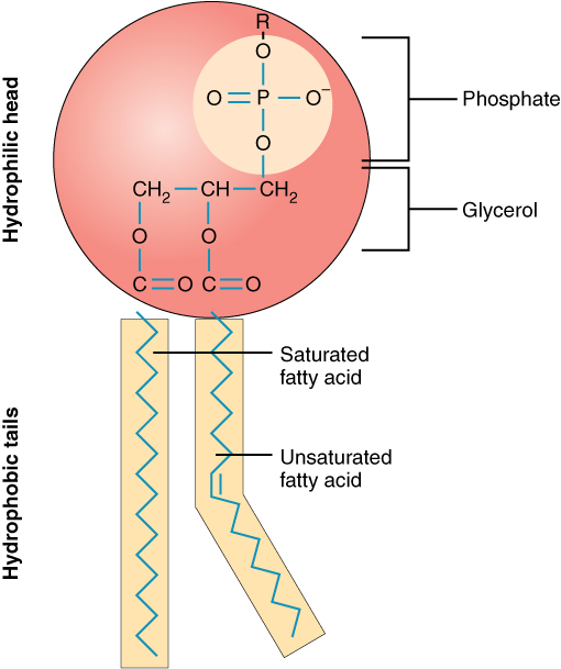
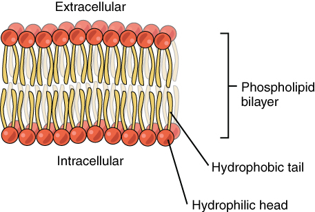
The plasma membrane is composed mainly of phospholipids, consisting of fatty acids and alcohol. The phospholipids in the plasma membrane are arranged in two layers, called a phospholipid bilayer. The simplified diagram in Figure 4.2.1 shows that each individual phospholipid molecule has a phosphate group head (in red) and two fatty acid tails (in yellow). The head “loves” water (hydrophilic), and the tails “hate” water (hydrophobic). The water-hating tails are on the interior of the membrane. In contrast, the water-loving heads point outward toward the cytoplasm (intracellular) or the fluid surrounding the cell (extracellular).
Hydrophobic molecules can easily pass through the plasma membrane if they are small enough because they are water-hating, like the interior of the membrane. Hydrophilic molecules, conversely, cannot pass through the plasma membrane — at least not without help — because they are water-loving like the membrane’s exterior.
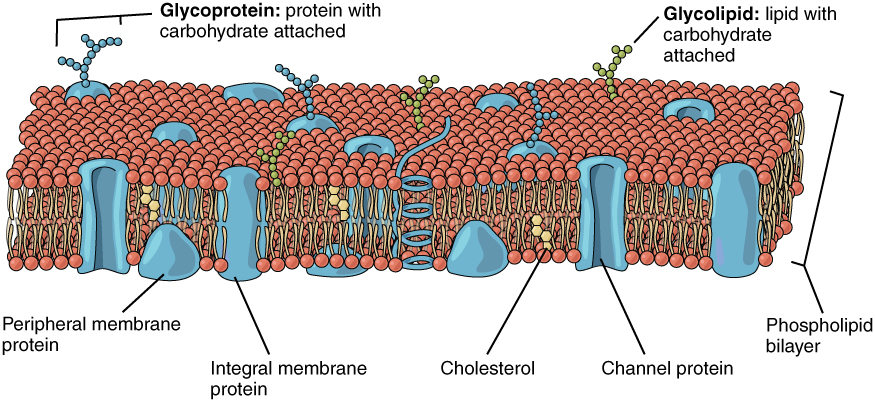
The plasma membrane also contains other molecules, primarily other lipids and proteins. For example, molecules of the steroid lipid cholesterol (shown in yellow in Figure 4.2.3) help the plasma membrane keep its shape. Proteins in the plasma membrane (shown blue) include transport proteins that assist other substances in crossing the cell membrane, receptors that allow the cell to respond to chemical signals in its environment, and cell-identity markers that indicate what type of cell it is and whether it belongs in the body. The plasma membrane is often described as a fluid mosaic, meaning the components can flow and change position while maintaining the basic integrity of the membrane.
The Cytoplasm
The cytoplasm is a thick, usually colourless solution that fills each cell and is enclosed by the cell membrane. Cytoplasm presses against the cell membrane, filling out the cell and giving it its shape. In eukaryotic cells, the cytoplasm includes all of the material inside the cell and outside the nucleus. All of the organelles in eukaryotic cells (such as the endoplasmic reticulum and mitochondria) are located in the cytoplasm. The cytoplasm helps to keep them in place. It is also the site of most metabolic activities in the cell, allowing materials to pass easily throughout the cell.
The portion of the cytoplasm surrounding organelles is called cytosol. Cytosol is the liquid part of the cytoplasm. It comprises about 80 percent water and contains dissolved salts, fatty acids, sugars, amino acids, and proteins (such as enzymes). These dissolved substances are needed to keep the cell alive and carry out metabolic processes. Enzymes dissolved in the cytosol, for example, break down larger molecules into smaller products that cell organelles can use. Waste products are also dissolved in the cytosol before they are taken in by vacuoles or expelled from the cell.
The Cytoskeleton
Although cytoplasm may appear to have no form or structure, it is actually highly organized. A framework of protein scaffolds called the cytoskeleton provides the cytoplasm and the cell with structure. The cytoskeleton consists of thread-like microfilaments, intermediate filaments, and microtubules crisscrossing the cytoplasm. As its name suggests, the cytoskeleton is like a cellular “skeleton.” It helps the cell maintain its shape and also helps to hold cell structures (like organelles) in place within the cytoplasm.
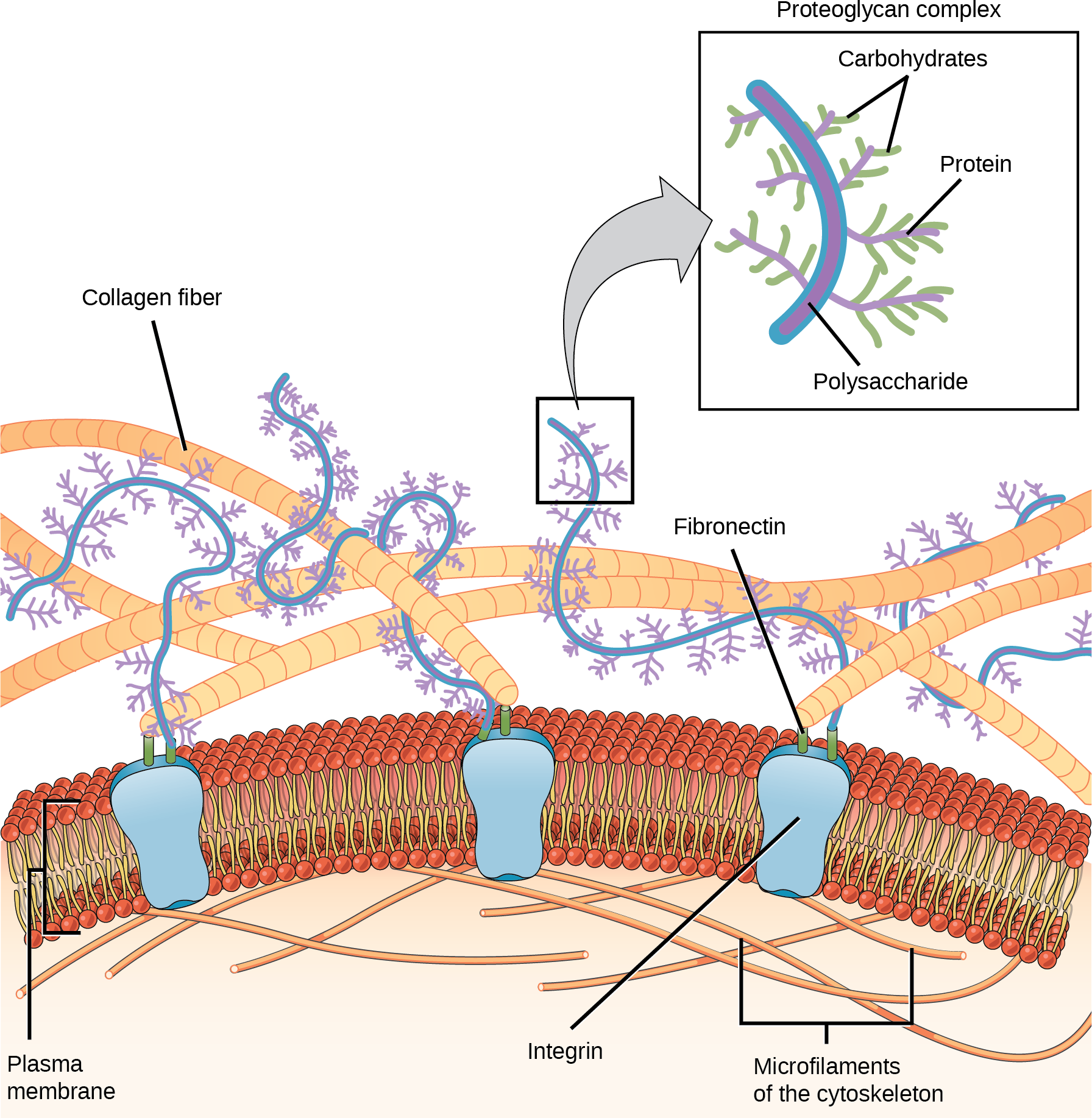
Extracellular Matrix
The extracellular matrix is a network of proteins and polysaccharides outside most animal cells. The extracellular matrix holds the cells together to form a tissue and allows the cells within the tissue to communicate with each other.
Cell Movement
Some cells need to be able to move to explore their surroundings, carry out vital functions, and react to external signals. The cytoskeleton plays a crucial role in facilitating movement by providing structural support and enabling changes in cell shape. Some cells, including some prokaryotes and some animal cells, have additional structures to help with movement.
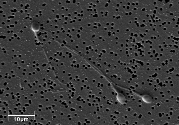
Flagella (singular flagellum) are long, whip-like structures that extend from the plasma membrane and help in movement. Sperm, Euglena, and many bacteria rely on flagella to help them swim through liquids. If present, a cell typically only has one or a few flagella.
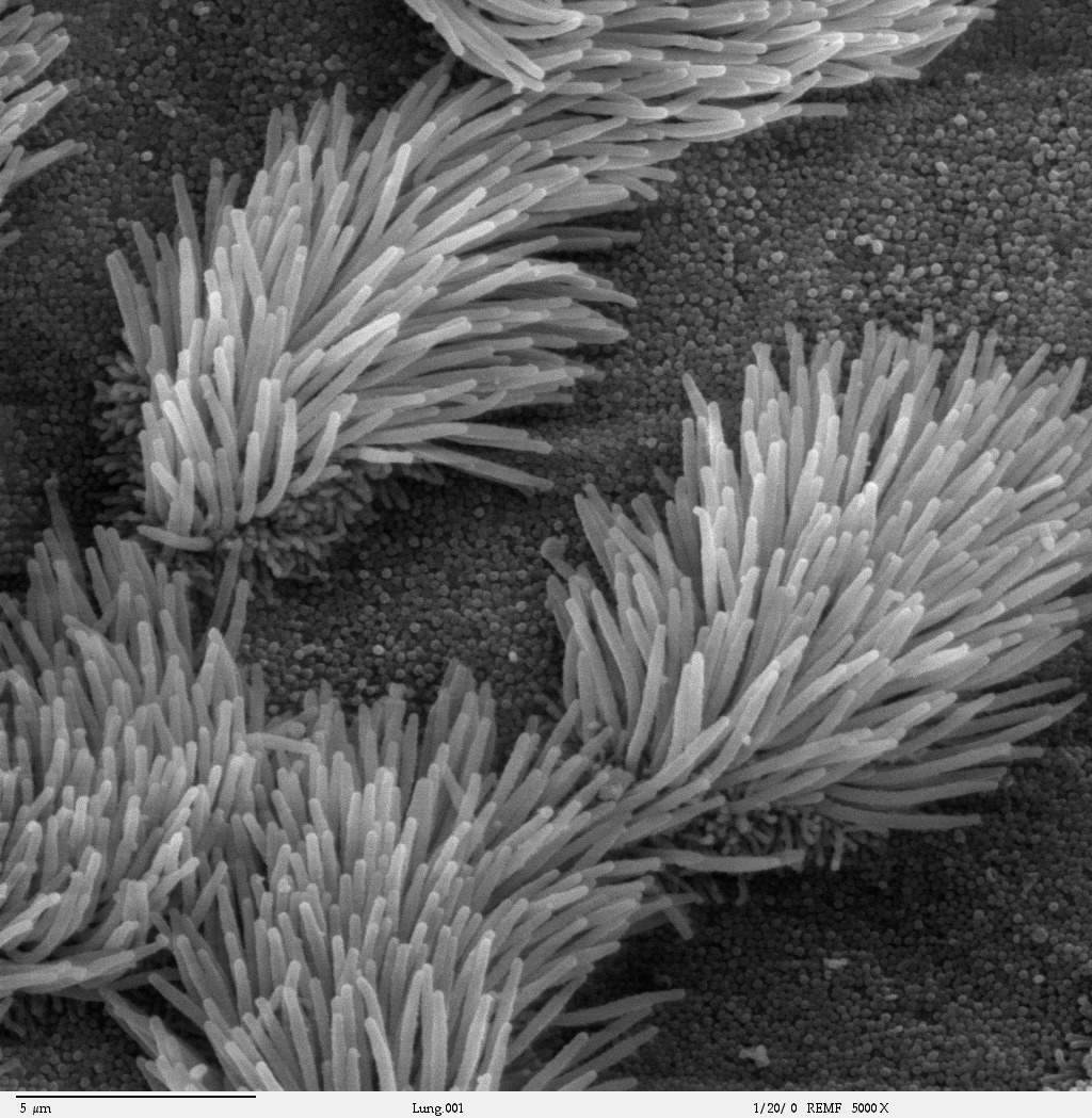
Cilia (singular cilium) are short, hair-like structures that help in movement. When cilia are present, they are many in number and extend along the entire plasma membrane surface. They can be used to move entire cells (such as paramecium) or move substances along the outer surface of the cell (for example, the cilia of cells lining the fallopian tubes that move the ovum toward the uterus or cilia lining the cells of the respiratory tract that sweep foreign particles and mucus toward the throat).
“4.4 Plasma Membrane” from Human Biology by Christine Miller is licensed under a Creative Commons Attribution-NonCommercial 4.0 International License, except where otherwise noted.
“4.5 Cytoplasm and Cytoskeleton” from Human Biology by Christine Miller is licensed under a Creative Commons Attribution-NonCommercial 4.0 International License, except where otherwise noted.
