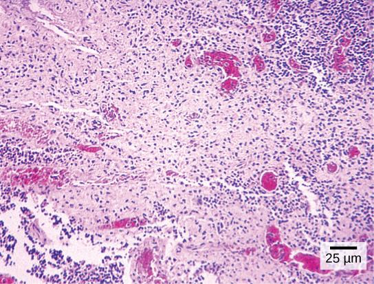
4.1 Cells
Your body has many kinds of cells, each specialized for a specific purpose. Just as a home is made from various building materials, the human body is constructed from many cell types. For example, epithelial cells protect the body’s surface, covering the organs and body cavities. Bone cells help to support and protect the body. Cells of the immune system fight invading bacteria. Red blood cells carry oxygen throughout the body. Each of these cell types plays a vital role during the body’s growth, development, and day-to-day maintenance. Despite their enormous variety, however, all cells share specific fundamental characteristics.
Figure 4.1.1 Diversity of human cells – a human T cell, developing nerve cells, a natural killer cell, red blood cells including T cells (orange) and platelets (green), and white blood cells.
Microscopy
Cells vary in size. With few exceptions, individual cells are too small to be seen with the naked eye, so scientists use microscopes to study them. A microscope is an instrument that magnifies an object. Most images of cells are taken with a microscope. Photographs taken with microscopes are called micrographs.
Light Microscopes
Light microscopes use visible light and a series of lenses to magnify specimens. They are relatively inexpensive, easy to use, and can provide detailed images of living organisms and cells. The downside is that they can only magnify up to around 1000 times, which may sound like a lot, but is still insufficient for observing small details or structures.

Electron Microscopes
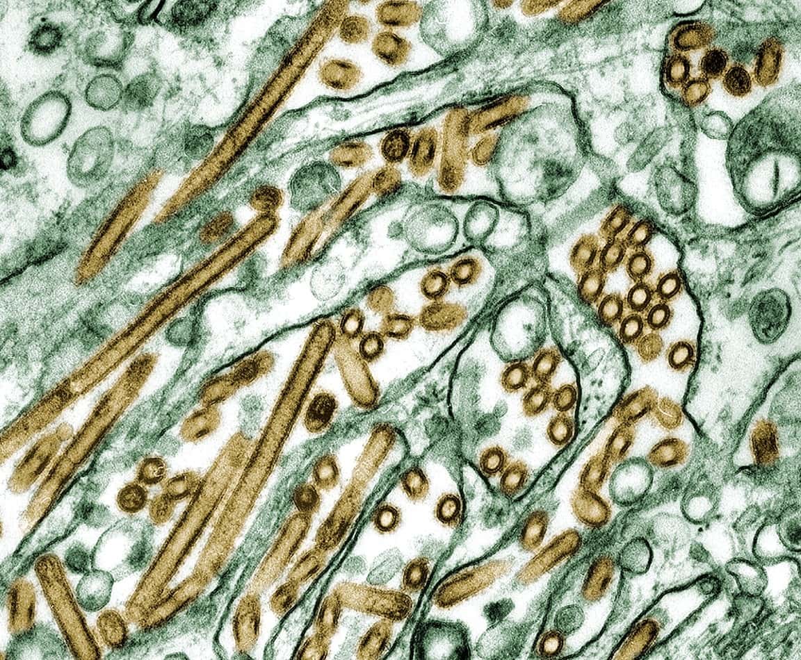
Electron microscopes use a beam of electrons instead of light to view specimens. They can magnify significantly smaller structures in much more detail than light microscopes. Scanning electron microscopes are used to see the details of cell surfaces. Transmission electron microscopes provide details of a cell’s internal structures. The downside is that electron microscopes are significantly more expensive and bulkier than light microscopes, so they are less easily accessible. The preparation to view a specimen under an electron microscope will also kill it, so these microscopes cannot be used to view live organisms.

Cell Theory
The first time the word cell was used to refer to these tiny units of life was in 1665 by a British scientist named Robert Hooke. Hooke was one of the earliest scientists to study living things under a microscope. The microscopes of his day were not very strong, but Hooke could still make an important discovery. When he looked at a thin slice of bark from a cork oak tree under his microscope, he was surprised to see that it was composed of many tiny units. Hooke called these units cells because they resembled cells in a monastery.
Soon after, Anton van Leeuwenhoek in Holland made other important discoveries using a microscope. Leeuwenhoek made his own microscope lenses, and he was so good at it that his microscope was more powerful than other microscopes of his day. In fact, Leeuwenhoek’s microscope was almost as strong as modern light microscopes. Using his microscope, Leeuwenhoek was the first person to observe human cells and bacteria.
By the early 1800s, scientists had observed cells of many different organisms. These observations led two German scientists, Theodor Schwann and Matthias Jakob Schleiden, to propose cells as the basic building blocks of all living things. Around 1850, a German doctor named Rudolf Virchow was studying cells under a microscope when he saw them dividing and forming new cells. He realized that living cells produce new cells through division. Virchow proposed that living cells arise only from other living cells based on this realization. The ideas of all three scientists — Schwann, Schleiden, and Virchow — led to cell theory, which is one of the fundamental theories unifying all of biology.
Cell theory states that:
- All organisms are made of one or more cells.
- All the life functions of organisms occur within cells.
- All cells come from existing cells.
Categories of Cells
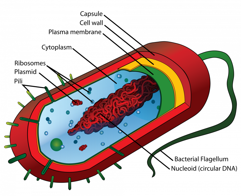
All cells share four common components:
- A plasma membrane, an outer covering that separates the cell’s interior from its surrounding environment.
- Cytoplasm, consisting of a jelly-like region within the cell in which other cellular components are found.
- DNA, the cell’s genetic material.
- Ribosomes, particles that synthesize proteins.
Based on whether or not they have a nucleus, there are two basic types of cells: prokaryotic cells and eukaryotic cells.
Prokaryotic Cells
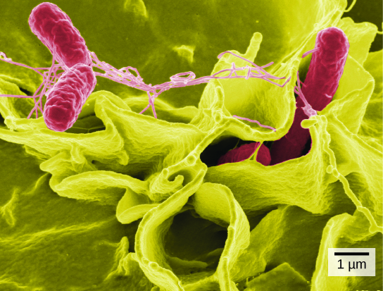
Prokaryotic cells are cells without a nucleus. The DNA in prokaryotic cells is in the cytoplasm in a region called the nucleoid, rather than enclosed within a nuclear membrane. In addition, these cells are typically smaller than eukaryotic cells and contain fewer organelles. Prokaryotic cells are found in single-celled organisms, such as the bacterium represented by the model in Figure 4.3.3. Organisms with prokaryotic cells are called prokaryotes. They were the first type of organisms to evolve and are still the most common.
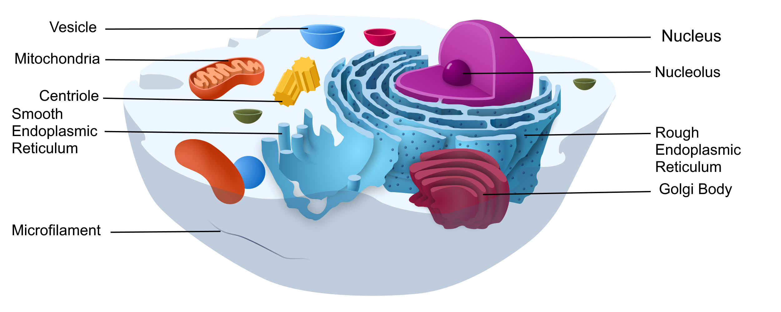
Eukaryotic Cells
Eukaryotic cells are cells that contain a nucleus. Eukaryotic cells are usually 10-100 times larger than prokaryotic cells. They are found in some single-celled and multicellular organisms. Organisms with eukaryotic cells are called eukaryotes, and they range from fungi to humans.
Besides a nucleus, eukaryotic cells also contain other organelles. An organelle is a structure within the cytoplasm that performs a specific job in the cell. Organelles called mitochondria, for example, provide energy to the cell, and organelles called vesicles store substances in the cell. Organelles allow eukaryotic cells to carry out more functions than prokaryotic cells can.
Comparing Prokaryotic and Eukaryotic Cells
Table 4.1.1 Comparing Prokaryotic Cells and Eukaryotic Cells (Simon, E. J., Dickey, J. 2018, pg 57)
| Prokaryotic Cells | Eukaryotic Cells |
|---|---|
| First evolved approximately 3.5 billion years ago | First evolved approximately 2.1 billion years ago |
| Found in bacteria and archaea | Found in protists, plants, fungi, and animals |
| Smaller, simpler | Larger, more complex |
| Most have cell walls; some have capsules, fimbriae, and/or flagella | Plant cells have cell walls; animal cells are surrounded by an extracellular matrix |
| Have a plasma membrane | Have a plasma membrane |
| No membrane-bound organelles | Membrane-bound organelles (for example, nucleus, ER) |
| Have a nucleoid region containing a single circular chromosome | Have a nucleus containing one or more linear chromosomes |
| Have ribosomes | Have ribosomes |
Cell Size
Most organisms, even very large ones, have microscopic cells. Why don’t cells get bigger instead of remaining tiny and multiplying? Why aren’t you one giant cell rolling around school? What limits cell size?
Once you know how a cell functions, the answers to these questions are clear. To carry out life processes, a cell must be able to quickly pass substances in and out of the cell. For example, it must be able to pass nutrients and oxygen into the cell and waste products out of the cell. Anything that enters or leaves a cell must cross the plasma membrane. The size of a cell is limited by its need to pass substances across the plasma membrane. Small cells maintain a proper surface area-to-volume ratio. Large cells have too much volume compared to surface area so are not as efficient at transport processes.
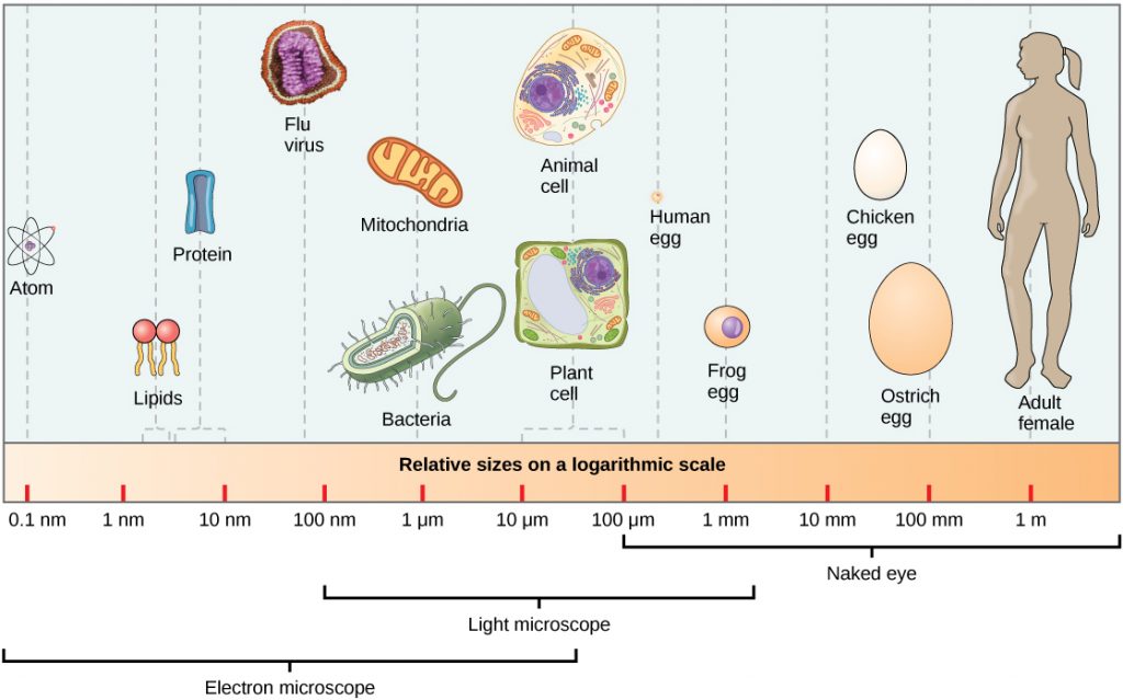
Image Description
The following can be seen in an electron microscope and/or light microscope: Atom, Lipids, Protein, Flu virus, Mitochondria, Bacteria. Animal Cell. The following can be seen by the naked eye: Plant Cell, Human egg, Frog egg, Chicken egg, Ostrich egg, Adult human.
“4.2 Discovery of Cells and Cell Theory” from Human Biology by Christine Miller is licensed under a Creative Commons Attribution-NonCommercial 4.0 International License, except where otherwise noted.
“Chapter 3: Introduction to Cell Structure and Function” from Biology and the Citizen by Colleen Jones is licensed under a Creative Commons Attribution 4.0 International License, except where otherwise noted.
“4.3 Variation in Cells” from Human Biology by Christine Miller is licensed under a Creative Commons Attribution-NonCommercial 4.0 International License, except where otherwise noted.
