7.3 Mitosis and Cytokinesis
The mitotic phase is the part of the eukaryotic cell cycle when the cell is dividing. It is a multistep process during which the duplicated chromosomes are aligned, separated, and moved to opposite poles of the cell. Then, the cell is divided into two new identical daughter cells. The mitotic phase consists of two overlapping processes: mitosis and cytokinesis.
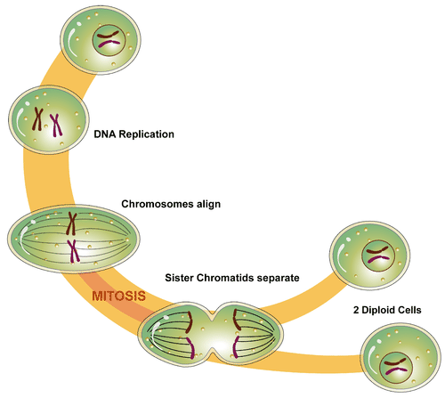
Figure 7.3.1 Description
The image is a diagram illustrating the process of mitosis, the type of cell division that results in two genetically identical diploid daughter cells.
- Starting Cell (Interphase): The diagram begins with a single diploid cell containing a nucleus with uncondensed chromatin.DNA Replication occurs, and chromosomes duplicate, each consisting of two sister chromatids.
- Chromosomes Align (Metaphase): The chromosomes, now condensed and visible, align at the center of the cell on the metaphase plate. The spindle fibres are attached to the centromeres of the chromosomes.
- Sister Chromatids Separate (Anaphase-Telophase): The sister chromatids are pulled apart by spindle fibres and move towards opposite poles of the cell. The cell starts to elongate, and the nuclear envelope begins to reform.
- Formation of Two Diploid Cells (Cytokinesis): The cell membrane pinches in, leading to the formation of two genetically identical diploid daughter cells. Each daughter cell has the same chromosome number as the original parent cell.
Mitosis
Mitosis is the process in which the nucleus of a eukaryotic cell divides. During mitosis, the two sister chromatids that make up each chromosome separate from each other and move to opposite poles of the cell. This type of cell division begins with one diploid cell and results in two identical diploid daughter cells. Mitosis is used for growth and replacing dead or damaged cells. It can also be used for asexual reproduction in some organisms.
Mitosis occurs in four phases: prophase, metaphase, anaphase, and telophase. Expand each phase below to learn more.
Phase 1: Prophase
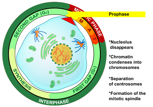
The first and longest phase of mitosis is prophase. During this phase, chromatin condenses into distinct chromosomes, each consisting of two sister chromatids joined at the centromere. Centrosomes move to opposite ends of the cell, initiating the formation of the spindle fibers. These fibers, composed of microtubules, are crucial for the subsequent steps of mitosis. The centrosomes play a key role in ensuring that the new cells formed after cell division contain a complete set of chromosomes. The nuclear envelope breaks down, allowing the spindle fibers to attach to the chromosomes.
Phase 2: Metaphase
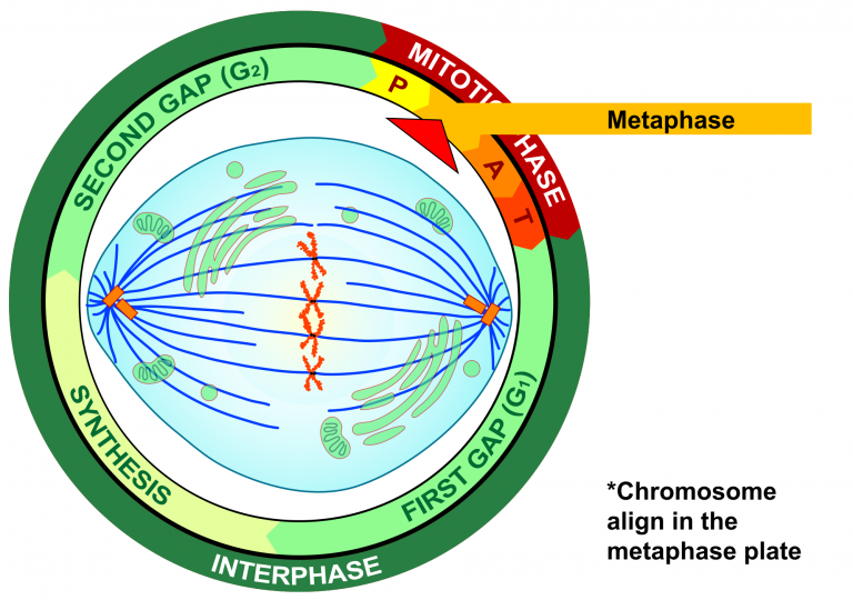
During metaphase, spindle fibers from opposite poles attach to the centromere of each chromosome, creating a “tug of war” effect. This tension ensures chromosomes align at the cell’s equator, known as the metaphase plate. The spindle fibres ensure that sister chromatids separate and go to different daughter cells when the cell divides.
Phase 3: Anaphase
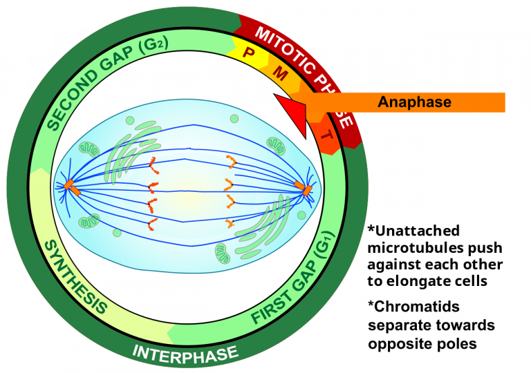
During anaphase, sister chromatids are pulled apart by the spindle fibers. Each chromatid, now an individual chromosome, moves toward opposite poles of the cell. This separation ensures that each new cell will receive an identical set of chromosomes. Spindle fibers not attached to chromosomes elongate, pushing the poles further apart and helping to stretch the cell.
Phase 4: Telophase
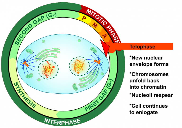
During telophase, the separated chromosomes reach the opposite poles of the cell. Telophase is the reverse of prophase. The spindle fibers begin to disassemble, and the nuclear envelope re-forms around each set of chromosomes, creating two distinct nuclei. The chromosomes start to decondense back into chromatin. Cytokinesis often starts during this phase, to split the cytoplasm into two daughter cells.
Cytokinesis
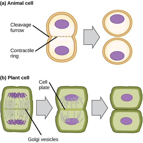
Cytokinesis is the second part of the mitotic phase, during which cell division is completed by the physical separation of the cytoplasmic components into two daughter cells. Although the stages of mitosis are similar for most eukaryotes, the process of cytokinesis is quite different for eukaryotes with cell walls, such as plant cells.
In cells such as animal cells that lack cell walls, cytokinesis begins following the onset of anaphase. A contractile ring composed of actin filaments forms inside the plasma membrane at the former metaphase plate. The actin filaments pull the cell’s equator inward, forming an indentation called the cleavage furrow. The furrow deepens as the actin ring contracts, and eventually, the membrane and cell are split in two (Figure 7.3.6).
A cleavage furrow is impossible in plant cells because of the rigid cell walls surrounding the plasma membrane. A new cell wall must form between the daughter cells. During interphase, the Golgi apparatus accumulates enzymes, structural proteins, and glucose molecules prior to breaking up into vesicles and dispersing throughout the dividing cell. These Golgi vesicles move on microtubules to collect at the metaphase plate during telophase. There, the vesicles fuse from the centre toward the cell walls; this structure is called a cell plate. As more vesicles fuse, the cell plate enlarges until it merges with the cell wall at the cell’s periphery. Enzymes use the glucose that has accumulated between the membrane layers to build a new cell wall of cellulose. The Golgi membranes become the plasma membrane on either side of the new cell wall (Figure 7.3.6).
Exercise 7.3.1
Text Description
Drag to arrange the images in the correct sequence
- An image illustrating telophase and cytokinesis
- An image illustrating anaphase
- An image illustrating metaphase
- An image illustrating prophase
Answers:
-
- An image illustrating prophase
- An image illustrating metaphase
- An image illustrating anaphase
- An image illustrating telophase and cytokinesis
Text Description
“4.13 Mitosis and Cytokinesis” from Human Biology by Christine Miller is licensed under a Creative Commons Attribution-NonCommercial 4.0 International License, except where otherwise noted.
“6.2 The Cell Cycle” from Biology and the Citizen by Colleen Jones is licensed under a Creative Commons Attribution 4.0 International License, except where otherwise noted.
